
Introduction
Ringworm, also known as dermatophytosis, is a superficial fungal infection of the skin, hair, and nails caused by dermatophytes — fungi that feed on keratin. Despite its name, ringworm is not caused by a worm, but by fungi belonging mainly to the genera Microsporum, Trichophyton, and Epidermophyton. It affects a wide range of domestic animals, especially cats, dogs, cattle, and horses, and is highly zoonotic, meaning it can be transmitted to humans.
Etiology and Causative Agents
- Genus: Microsporum, Trichophyton, Epidermophyton.
- Most common veterinary species:
- Microsporum canis (cats, dogs, zoonotic).
- Microsporum gypseum (soil-associated).
- Trichophyton mentagrophytes (dogs, rodents, humans).
- Reservoirs: Infected animals, contaminated environment (spores on bedding, grooming tools, soil).
Morphology and Resistance
- Fungi are filamentous, producing septate hyphae and asexual spores (arthroconidia).
- Spores can survive for months to years in the environment, making eradication difficult.
- Resistant to desiccation but sensitive to disinfectants such as bleach, iodine compounds, and enilconazole.
Transmission and Epidemiology
Ringworm spreads by:
- Direct contact with infected animals or humans.
- Indirect contact with contaminated objects (brushes, collars, bedding).
- Environmental exposure to fungal spores in soil or surfaces.
Risk groups: Young animals, immunosuppressed individuals, overcrowded shelters, and farms.
Pathogenesis and Clinical Signs
Dermatophytes invade keratinized tissues (stratum corneum, hair, nails) but do not penetrate deeper tissues.
The infection causes an inflammatory reaction in the skin, leading to:
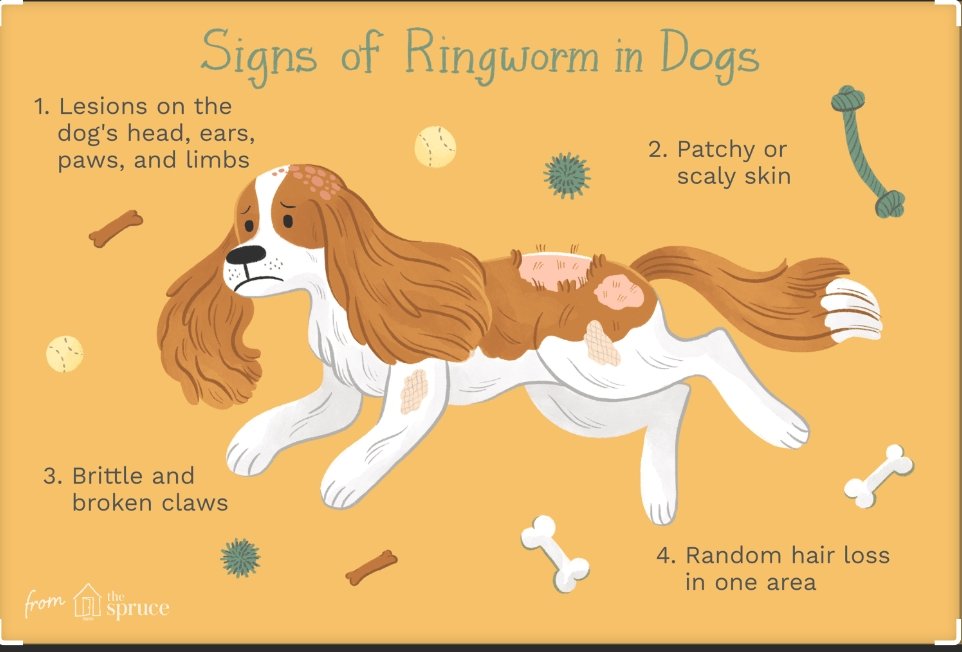
In animals:
- Circular areas of alopecia (hair loss).
- Scaly, crusty, or inflamed lesions with a characteristic ring-like appearance.
- Broken hairs and brittle coat.
- Itching may or may not be present.
- In cattle: “Trichophytosis” with grayish-white plaques on the head and neck.
In humans:
- Itchy, ring-shaped red lesions on skin (classic “ringworm” appearance).
- On scalp: patchy hair loss (tinea capitis).
- On body: red, scaly rings (tinea corporis).
Incubation Period
- Typically 1–3 weeks after exposure, depending on fungal load and host immunity.
Laboratory Diagnosis
Accurate diagnosis is essential since ringworm resembles other skin diseases. Methods include:
- Wood’s lamp examination: Some Microsporum species fluoresce apple-green.
- Microscopy: KOH mount of hair/skin scrapings showing hyphae and arthroconidia.
- Culture: On Sabouraud’s dextrose agar with antibiotics, colonies of dermatophytes appear within 1–3 weeks.
- PCR: Molecular confirmation in reference laboratories.
Prevention and Control
- Environmental decontamination: Regular disinfection with bleach, enilconazole, or other antifungal agents.
- Isolation of infected animals to prevent spread in households or shelters.
- Hygiene: Hand washing, wearing gloves when handling infected pets.
- Regular grooming and inspection of pets and livestock.
- Vaccination: In some countries, vaccines against Trichophyton verrucosum (cattle) and Microsporum canis (cats/dogs) are available to aid prevention.
Treatment
- Topical antifungals: Miconazole, enilconazole, clotrimazole creams or sprays.
- Systemic antifungals (for severe or generalized cases): Itraconazole, terbinafine, griseofulvin (in animals, under veterinary supervision).
- Shampoos and dips (lime sulfur, miconazole-chlorhexidine).
- Environmental cleaning: Repeated disinfection to eliminate spores.
Importance for Veterinary and Public Health
Ringworm is a highly contagious zoonotic disease, especially important in households with children, the elderly, or immunocompromised people. In farm animals, it also causes economic losses due to reduced leather quality and market restrictions.
At Dr. Boudi Widad Veterinary Clinic, we provide:
- Accurate diagnostic testing for rapid identification of dermatophytes.
- Comprehensive treatment plans combining topical and systemic therapy.
- Preventive guidance for pet owners, shelters, and farmers to minimize reinfection.